Understanding fetal biometry, specifically ac fl hc bpd, is essential in prenatal care. Gestational age assessment relies heavily on these measurements, providing crucial insights into fetal development. The Sonography Department at the University Hospital utilizes these parameters routinely. Further, research by prominent figure Dr. Jane Doe, emphasizes the role of ac fl hc bpd in identifying potential growth abnormalities. The Hadlock formula, a widely used tool, incorporates ac fl hc bpd to estimate fetal weight and monitor growth trends.
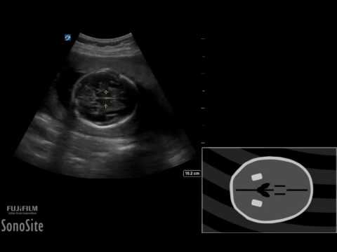
Image taken from the YouTube channel Sonosite , from the video titled How To: Pregnancy BPD HC AC and FL Measurements 3D Video .
Understanding Fetal Measurements During Pregnancy: A Guide to AC, FL, HC, and BPD
The journey of pregnancy is a remarkable process, filled with anticipation and wonder. From the moment of conception, a complex series of developmental milestones unfold within the womb. Prenatal care, a cornerstone of modern obstetrics, provides crucial monitoring and support throughout this transformative period.
One of the most common and informative tools used in prenatal care is ultrasound imaging.
Ultrasound allows healthcare providers to visualize the developing fetus, assess its growth, and identify potential concerns.
The Role of Ultrasound in Fetal Development Monitoring
Ultrasound technology uses sound waves to create images of the fetus in utero. These images allow doctors and sonographers to monitor the baby’s progress, estimate gestational age, and screen for certain developmental conditions.
Regular prenatal ultrasounds offer invaluable insights into the baby’s well-being and can help guide medical decisions throughout the pregnancy.
Unveiling AC, FL, HC, and BPD: Key Indicators of Fetal Well-being
During an ultrasound, sonographers take specific measurements of the fetus, including:
- AC (Abdominal Circumference): The measurement around the baby’s abdomen.
- FL (Femur Length): The length of the femur, the long bone in the thigh.
- HC (Head Circumference): The measurement around the baby’s head.
- BPD (Biparietal Diameter): The distance between the two sides of the baby’s head.
These measurements, often referred to as fetal biometry, are vital indicators of fetal growth and development.
They provide valuable information about the baby’s gestational age, overall size, and potential health concerns.
Why These Measurements Matter
AC, FL, HC, and BPD are carefully assessed and compared to established growth charts to determine if the fetus is developing within a healthy range.
These measurements can help identify potential growth abnormalities, such as intrauterine growth restriction (IUGR) or macrosomia (excessively large baby).
They also play a critical role in estimating fetal weight, which is important for planning delivery.
Acknowledging Variations: Understanding the Range of "Normal"
It’s important to remember that fetal growth is not an exact science. Variations in measurements are common and don’t necessarily indicate a problem.
Each baby develops at its own pace, and factors such as genetics, maternal health, and even the baby’s position during the ultrasound can influence the measurements.
Healthcare providers consider these factors when interpreting the results and will conduct further evaluation if there are any concerns.
Your Guide to Understanding Fetal Measurements
This guide aims to provide you with a clear and accessible understanding of AC, FL, HC, and BPD.
By offering context and clarifying the significance of these measurements, we hope to empower you to navigate your prenatal journey with greater confidence and understanding.
We will delve into each measurement individually, explain how they are obtained, and discuss their implications for fetal well-being.
Our goal is to provide you with the knowledge you need to engage in informed conversations with your healthcare provider and actively participate in your prenatal care.
The information gleaned from these glimpses inside the womb are more than just images; they are data points that, when carefully analyzed, paint a detailed picture of the developing baby’s health and progress. Let’s delve into the specific measurements that comprise fetal biometry, understanding what they signify and how they are obtained.
Decoding Fetal Biometry: A Deep Dive into Core Measurements
Fetal biometry is the systematic measurement of a fetus’s physical dimensions using ultrasound technology. It is a cornerstone of prenatal care, providing critical information about a baby’s growth, development, and gestational age.
These measurements are meticulously obtained by trained sonographers and interpreted by physicians to assess the overall health and well-being of the fetus. Let’s examine the key components of fetal biometry: Abdominal Circumference (AC), Femur Length (FL), Head Circumference (HC), and Biparietal Diameter (BPD).
What is Fetal Biometry?
Fetal biometry involves a series of standardized measurements of the fetus taken during ultrasound examinations. These measurements typically include AC, FL, HC, and BPD, though others may be used depending on the gestational age and clinical context.
The data collected through biometry is compared against established norms for gestational age. This comparison helps healthcare providers to assess whether the fetus is growing at an appropriate rate.
Fetal biometry is crucial for:
- Estimating gestational age, particularly in early pregnancy.
- Monitoring fetal growth patterns throughout the pregnancy.
- Identifying potential growth abnormalities, such as intrauterine growth restriction (IUGR) or macrosomia (excessively large baby).
- Calculating estimated fetal weight (EFW).
AC (Abdominal Circumference)
Definition and Significance of AC
Abdominal Circumference (AC) measures the distance around the fetus’s abdomen. It is a crucial indicator of fetal size and nutritional status.
AC reflects the size of the fetal liver, spleen, and subcutaneous fat, making it particularly sensitive to changes in fetal nutrition. A disproportionately small AC can be a sign of IUGR, while a larger-than-expected AC may suggest gestational diabetes in the mother or fetal macrosomia.
How AC is Measured During Ultrasound
During an ultrasound, the sonographer obtains the AC measurement by carefully positioning the ultrasound transducer to capture a cross-sectional image of the fetal abdomen. Specific anatomical landmarks, such as the umbilical vein and stomach, are used to ensure accurate and consistent measurements.
The sonographer then uses electronic calipers on the ultrasound machine to trace the circumference of the abdomen on the image.
What AC Tells Us About Fetal Development and Nutrition
AC provides valuable insight into fetal growth and nutrition. A normal AC indicates that the fetus is receiving adequate nutrients from the mother and is growing at an appropriate rate.
Serial AC measurements, taken over time, can reveal patterns of growth and help identify potential problems early on.
AC, when combined with other biometric measurements, contributes to a more complete picture of fetal well-being.
FL (Femur Length)
Definition and Significance of FL
Femur Length (FL) is the measurement of the longest bone in the body. It is found in the thigh, extending from the hip to the knee.
FL is a reliable indicator of gestational age, particularly during the second trimester. It also contributes to estimating fetal size and detecting skeletal dysplasia.
How FL is Measured
The sonographer identifies the femur on the ultrasound image. They then measure its length from one end to the other using electronic calipers.
Care is taken to measure the entire length of the ossified (bony) portion of the femur, excluding the cartilaginous ends.
Importance in Determining Gestational Age
FL is most accurate for determining gestational age between 14 and 22 weeks of gestation. After this point, the variability in FL increases, and other measurements like HC and BPD may be more reliable for gestational age estimation.
FL is particularly useful when the date of the last menstrual period is unknown or uncertain.
HC (Head Circumference)
Definition and Importance of HC
Head Circumference (HC) measures the distance around the fetus’s head. It’s an important parameter for assessing fetal growth and brain development.
HC is less affected by nutritional factors than AC. Therefore, it can be a more reliable indicator of gestational age, especially in cases of suspected IUGR.
Measurement Process for HC
The sonographer obtains the HC measurement by positioning the ultrasound transducer to capture an axial view of the fetal head. This view shows specific anatomical landmarks, such as the thalami and cavum septum pellucidum.
Electronic calipers are then used to trace the circumference of the head. Precision and standardized technique are key to ensuring accuracy.
Relationship to Brain Development
HC is closely related to brain development. Significant deviations from the norm can raise concerns about potential neurological abnormalities, such as microcephaly (abnormally small head) or hydrocephalus (fluid accumulation in the brain).
Monitoring HC growth over time can provide valuable information about the rate of brain development.
BPD (Biparietal Diameter)
Definition and Significance of BPD
Biparietal Diameter (BPD) measures the distance between the two parietal bones on either side of the fetal head.
BPD is one of the earliest biometric measurements used to estimate gestational age. It is most accurate during the first and second trimesters.
How BPD is Measured
To measure BPD, the sonographer captures an axial view of the fetal head. They then use electronic calipers to measure the distance between the outer edge of one parietal bone to the inner edge of the opposite parietal bone.
Early Indicator of Gestational Age
BPD is a reliable indicator of gestational age, particularly between 12 and 20 weeks. It can be used in conjunction with other measurements, such as crown-rump length (CRL) in the first trimester, to accurately determine the gestational age of the fetus.
As the pregnancy progresses, the accuracy of BPD for gestational age estimation decreases. This is due to increasing variability in head shape.
Decoding the individual measurements gives us valuable data, but the true power of fetal biometry lies in its ability to paint a comprehensive picture when these data points are considered together. Understanding how these measurements relate to each other, and to the overall context of the pregnancy, is crucial for informed prenatal care.
Interpreting the Measurements: Putting the Pieces Together
The Role of Gestational Age
Gestational age, the time elapsed since the first day of the woman’s last menstrual period, is the cornerstone of interpreting fetal biometry. It provides the framework against which all measurements are evaluated.
A measurement that is perfectly normal at 20 weeks could be cause for concern at 30 weeks, and vice versa.
Fetal growth is not linear; it accelerates and decelerates at different stages of pregnancy. Therefore, comparing fetal measurements to the expected range for the specific gestational age is essential for accurate assessment.
Using the wrong gestational age as a reference point can lead to misinterpretations and unnecessary anxiety.
Understanding Growth Charts and Percentiles
How Growth Charts Are Used
Growth charts are graphical representations of the distribution of fetal measurements at different gestational ages. They are constructed from large datasets of normal pregnancies.
These charts allow healthcare providers to visualize how an individual fetus’s measurements compare to the average and the expected range.
Measurements are plotted on the chart, and their position relative to the reference curves indicates whether they are within the normal range, above average, or below average.
Meaning of Percentiles for Each Measurement (AC, FL, HC, BPD)
Percentiles are used to express the relative position of a measurement within the distribution.
For instance, if a fetus’s abdominal circumference (AC) is at the 50th percentile, it means that 50% of fetuses at the same gestational age have a smaller AC, and 50% have a larger AC.
Similarly, an AC at the 10th percentile means the fetus’s AC is smaller than 90% of fetuses at the same gestational age.
It’s important to note that a fetus at the 50th percentile is considered to be at the "average" size for its gestational age.
Normal vs. Concerning Variations in Percentiles
While the "normal" range is generally considered to be between the 10th and 90th percentiles, this is not a rigid cutoff. A measurement slightly outside this range may not necessarily indicate a problem.
However, measurements consistently below the 10th percentile or above the 90th percentile warrant further investigation.
Additionally, a significant drop in percentile rank over time is more concerning than a single measurement outside the "normal" range. This is because it may indicate a change in the fetus’s growth trajectory.
Healthcare providers consider the overall clinical picture, including the mother’s health history and other ultrasound findings, when interpreting percentile rankings.
Estimated Fetal Weight (EFW)
The Estimated Fetal Weight (EFW) is a calculation that combines AC, FL, HC, and BPD to estimate the fetus’s weight in grams or pounds.
EFW is a valuable tool for assessing fetal growth and identifying potential problems such as growth restriction or macrosomia.
The formulas used to calculate EFW are based on statistical models that relate these measurements to actual birth weights.
While EFW is a helpful estimate, it is not a perfect predictor of birth weight. There can be variations of up to 10-15% between the EFW and the actual birth weight.
Potential Concerns: Macrosomia and Microcephaly
Variations in fetal measurements can sometimes indicate potential health concerns.
Macrosomia, meaning "large body," refers to a fetus that is significantly larger than average for its gestational age. It is often defined as an EFW above the 90th percentile or a birth weight of more than 4000 grams (8 pounds 13 ounces).
Macrosomia can increase the risk of complications during labor and delivery, such as shoulder dystocia.
Microcephaly, meaning "small head," refers to a condition where the fetal head circumference (HC) is significantly smaller than average for its gestational age. It is often defined as an HC below the 3rd percentile.
Microcephaly can be associated with underlying brain development issues and may indicate a need for further evaluation and monitoring.
Decoding the individual measurements gives us valuable data, but the true power of fetal biometry lies in its ability to paint a comprehensive picture when these data points are considered together. Understanding how these measurements relate to each other, and to the overall context of the pregnancy, is crucial for informed prenatal care. Let’s delve into what happens when these carefully gathered pieces don’t quite fit the expected mold, and how healthcare professionals navigate those situations.
Addressing Potential Concerns: Variations and What They Mean
While fetal biometry provides valuable insights, it’s important to remember that variations from the norm can occur. These variations, while sometimes concerning, don’t automatically indicate a problem. They often necessitate further investigation and careful monitoring. One such potential concern is Intrauterine Growth Restriction (IUGR). Understanding IUGR, its identification, and management are essential aspects of prenatal care. Open communication with your obstetrician plays a vital role in navigating these situations.
Understanding Intrauterine Growth Restriction (IUGR)
IUGR is a condition where a fetus doesn’t grow at the expected rate inside the womb. This can lead to the baby being smaller than usual at birth.
Definition and Causes of IUGR
IUGR is defined as a condition where the fetus’s estimated weight is below the 10th percentile for its gestational age. This means that the baby is smaller than 90% of other babies at the same stage of pregnancy.
Several factors can contribute to IUGR. These include:
- Maternal health conditions: Such as high blood pressure, diabetes, or heart disease.
- Placental problems: Issues with the placenta’s ability to provide adequate nutrients and oxygen to the fetus.
- Multiple pregnancies: Twins or triplets may compete for resources, leading to slower growth for one or more fetuses.
- Fetal abnormalities: Genetic or structural problems in the fetus can sometimes cause IUGR.
- Maternal lifestyle factors: Smoking, alcohol consumption, or drug use during pregnancy can increase the risk of IUGR.
How AC, FL, HC, and BPD Are Used to Identify IUGR
Fetal biometry plays a crucial role in identifying IUGR. Measurements like Abdominal Circumference (AC), Femur Length (FL), Head Circumference (HC), and Biparietal Diameter (BPD) are carefully assessed.
- A disproportionately small AC is often a key indicator of IUGR, as it reflects the size of the fetal liver and abdominal organs, which are particularly sensitive to nutrient deprivation.
- Deviations in FL, HC, and BPD, when considered in conjunction with AC, further contribute to the diagnosis.
- Healthcare providers use growth charts to track these measurements over time. They look for patterns of slowing growth that deviate from the expected curve.
Management and Potential Outcomes of IUGR
The management of IUGR depends on the severity of the condition and the gestational age of the fetus.
- Close monitoring is essential, including regular ultrasound scans to assess fetal growth and amniotic fluid levels.
- Doppler studies may be used to evaluate blood flow in the umbilical cord and fetal brain.
- In some cases, early delivery may be necessary to ensure the baby’s well-being, especially if there are signs of fetal distress.
Potential outcomes of IUGR can vary. Some babies with IUGR may experience:
- Breathing difficulties.
- Low blood sugar.
- Difficulty maintaining body temperature after birth.
- Long-term developmental problems in severe cases.
However, with proper management and care, many babies with IUGR thrive and develop normally.
The Importance of Open Communication with Obstetricians
Navigating potential concerns like IUGR can be daunting. Open communication with your obstetrician is essential for understanding the situation. This enables informed decisions about your care and your baby’s well-being.
Discussing Concerns and Questions with Your Doctor
Don’t hesitate to voice any concerns or questions you have with your doctor. They are your primary source of information and support during this time. Ask them to explain the ultrasound findings in detail.
Ensure you understand the implications of the measurements and what they mean for your baby. It is also vital to inquire about potential causes of the variations and the steps being taken to monitor the situation.
The Role of Additional Testing and Monitoring
In some cases, additional testing and monitoring may be necessary to assess the fetal condition further.
- This may include more frequent ultrasound scans.
- Doppler studies to evaluate blood flow.
- In some instances, genetic testing to rule out underlying fetal abnormalities.
Your doctor will explain the purpose of these tests and what to expect. Being informed helps alleviate anxiety and empowers you to actively participate in your care.
The Role of Ultrasound in Monitoring Fetal Development
Ultrasound plays a pivotal role in monitoring fetal development and identifying potential concerns. Follow-up ultrasound scans are often recommended to track fetal growth and assess the overall health of the pregnancy.
Frequency and Purpose of Follow-Up Ultrasound Scans
The frequency of follow-up ultrasound scans will depend on the specific circumstances of your pregnancy.
- In cases of suspected IUGR, scans may be scheduled every few weeks to closely monitor fetal growth.
- These scans allow healthcare providers to assess whether the fetus is continuing to grow. Also to evaluate the amniotic fluid levels, and monitor blood flow.
Working with Obstetricians for Optimal Patient Care
Optimal patient care is achieved through a collaborative approach between you and your obstetrician. By actively participating in your care, asking questions, and voicing concerns, you can work together to ensure the best possible outcome for you and your baby. Trust in your doctor’s expertise. Don’t hesitate to seek a second opinion if you feel it’s necessary. Remember that you are an integral part of the care team.
Resources and Support: Navigating Your Prenatal Journey
Fetal biometry and the complexities of prenatal care can feel overwhelming. It’s natural to seek additional information and support as you navigate this journey. Remember, knowledge is power, and accessing reliable resources can empower you to make informed decisions about your and your baby’s health.
This section aims to provide you with a curated list of trustworthy online resources, guidance on finding supportive communities, and a framework for formulating insightful questions to ask your healthcare provider.
Reliable Online Resources: Your Digital Toolkit
The internet is awash with information, but not all sources are created equal. When researching prenatal topics, it’s crucial to rely on reputable organizations and evidence-based websites. Look for websites affiliated with medical societies, academic institutions, or government health agencies.
-
The American College of Obstetricians and Gynecologists (ACOG): ACOG offers comprehensive information on all aspects of pregnancy, childbirth, and women’s health. Their website features articles, FAQs, and patient education resources reviewed by medical experts.
-
The Society for Maternal-Fetal Medicine (SMFM): SMFM is a professional organization for physicians specializing in high-risk pregnancies. Their website provides access to research articles, clinical guidelines, and patient fact sheets.
-
The Centers for Disease Control and Prevention (CDC): The CDC offers reliable information on pregnancy-related health risks, vaccinations, and other essential topics.
-
The National Institutes of Health (NIH): NIH conducts and supports medical research. Their website provides access to research findings and publications on various pregnancy-related conditions.
When evaluating online information, consider the source’s credentials, the date of publication (ensure it’s current), and whether the information is supported by scientific evidence. Be wary of websites promoting unproven treatments or offering medical advice without proper qualifications.
Support Groups: Connecting with Other Parents
Pregnancy can be a deeply personal experience, but it’s also a shared one. Connecting with other expectant parents can provide invaluable emotional support, practical advice, and a sense of community.
Support groups offer a safe and confidential space to share your experiences, ask questions, and learn from others who understand what you’re going through. These groups can be found online or in person, through hospitals, birthing centers, or community organizations.
-
Online Forums and Communities: Numerous online forums and social media groups cater to expectant parents. These platforms offer a convenient way to connect with others from around the world, share your experiences, and ask questions. However, be mindful of the information shared in these forums, and always consult with your healthcare provider before making any medical decisions.
-
Local Support Groups: Many hospitals and birthing centers offer prenatal support groups led by trained facilitators. These groups provide a structured environment for learning about pregnancy, childbirth, and parenting, and for connecting with other local parents.
-
Specialized Support Groups: If you’re facing specific challenges, such as a high-risk pregnancy or a diagnosis of IUGR, consider joining a specialized support group. These groups offer a more focused and tailored approach to addressing your unique needs and concerns.
Questions to Ask Your Doctor: Empowering Informed Decisions
Your obstetrician is your primary source of information and guidance throughout your pregnancy. Don’t hesitate to ask questions and express your concerns. There are no "silly" questions when it comes to your health and your baby’s well-being.
Preparing a list of questions before each appointment can help you make the most of your time with your doctor. Here are some questions you might consider asking related to fetal biometry:
- What do my baby’s measurements mean in the context of my overall pregnancy?
- Are my baby’s measurements within the normal range for my gestational age?
- If there are variations, what could be the potential causes?
- What additional testing or monitoring is recommended, and why?
- What are the potential outcomes based on my baby’s growth patterns?
- What steps can we take to optimize my baby’s growth and development?
- How will these measurements influence our birth plan?
Remember that proactive communication is key to ensuring the best possible care for you and your baby. By staying informed, connecting with others, and asking thoughtful questions, you can navigate your prenatal journey with confidence and peace of mind.
FAQs: Understanding AC FL HC BPD
Here are some frequently asked questions to help clarify AC FL HC BPD (Abdominal Circumference, Femur Length, Head Circumference, and Biparietal Diameter) as discussed in our guide.
What exactly does AC FL HC BPD measure?
AC FL HC BPD refers to key fetal measurements obtained during prenatal ultrasounds. AC is the Abdominal Circumference, FL is the Femur Length, HC is the Head Circumference, and BPD represents the Biparietal Diameter (head width). These measurements help assess fetal growth and development.
Why are AC FL HC BPD measurements important during pregnancy?
These measurements, AC FL HC BPD, are crucial for estimating gestational age, monitoring growth patterns, and detecting potential abnormalities. Comparing these values to standard growth charts allows doctors to identify if a baby is growing too slowly, too quickly, or if further investigation is warranted.
What if my baby’s AC FL HC BPD measurements are outside the normal range?
If any of the AC FL HC BPD measurements are outside the typical range for your gestational age, your doctor will likely recommend further evaluation. This might include additional ultrasounds, genetic testing, or other assessments to determine the cause and any necessary interventions. Remember that slight variations are common and don’t always indicate a problem.
Where can I find more in-depth information about AC FL HC BPD and fetal development?
Our guide includes links to several resources, including medical journals, reputable websites, and organizations dedicated to prenatal health. We also suggest discussing any specific concerns about your baby’s AC FL HC BPD results with your healthcare provider for personalized guidance.
So, that’s a wrap on ac fl hc bpd! Hopefully, this has helped clear things up and given you a solid foundation. Best of luck in your continued learning!